
Effect of a Composition Containing Chitosan Gel and Signalling Molecules of the Extracellular Matrix on Healing of Purulent Wounds
We studied the effect of combined preparation on the basis of chitosan containing a new of the peptide complex isolated from extracellular space leaves of aloe vera at a concentration of 0,001 mcg/ml and belonging to the group of homeostatic signal molecules on healing of purulent wounds in mammals in vivo.

Fig. 1. Healing of skin wound on day 3. Hematoxylin and eosin staining. 1)inflammatory focus, 2) derma, 3) subcutaneous adipose tissue, 4) crust.
Here and in Figs. 2 and 3: a) wound infected with pus; b) wound without pus contamination; c) application of levomecol; d) application of chitosan gel containing S.M.E.M.®
Day 3
On day 3 after wound infliction, a non-healing wound without epithelium in the central zone was observed in all cases. Pronounced inflammation with neutrophilic infiltration was seen in the derma. The effects of the test preparations cannot be adequately evaluated at this stage.
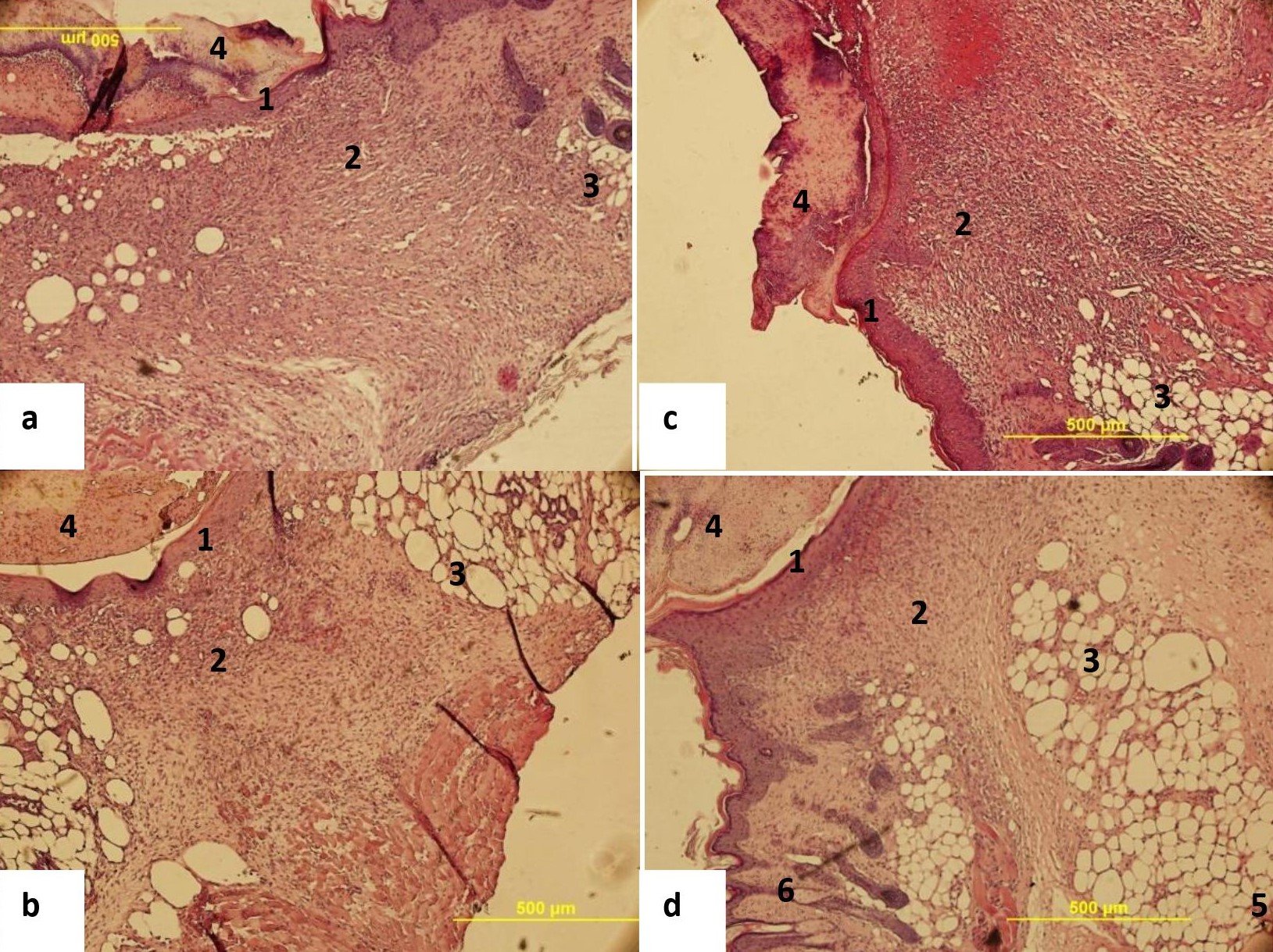
Fig. 2. Healing of skin wound on day 8. Hematoxylin and eosin staining. 1)epithelium, 2) derma, 3)subcutaneous adipose tissue, 4) crust, 5) gland ducts.
Day 8
On day 8 after wound infliction, we observed incomplete epithelization of infected wounds, the crust adherent to the wound surface, inflammatory focus and numerous blood vessels in the derma, formation of fibrous scar, and poorly developed adipose tissue (Fig.2,a). In the wound not contaminated with pus, epithelization and crust separation from the epithelium were observed. In the derma, the formation of fibrous scar, minor inflammatory foci, and well-developed subcutaneous adipose tissue with gland ducts were seen (Fig.2,b). In purulent wound treated with levomecol, pronounced inflammatory reaction was observed in the derma. Epithelization was not completed and the crust tightly adhered to the wound; poorly developed adipose tissue and the formation of fibrous scar were observed (Fig.2,c). Application of the chitosan-based gel containing S.M.E.M.® on the wound led to its intensive epithelization and complete crust detachment. No inflammatory foci were seen in the derma, the formation of gland ducts and muscle elements was seen in the subcutaneous adipose tissue. (Fig.2,d).
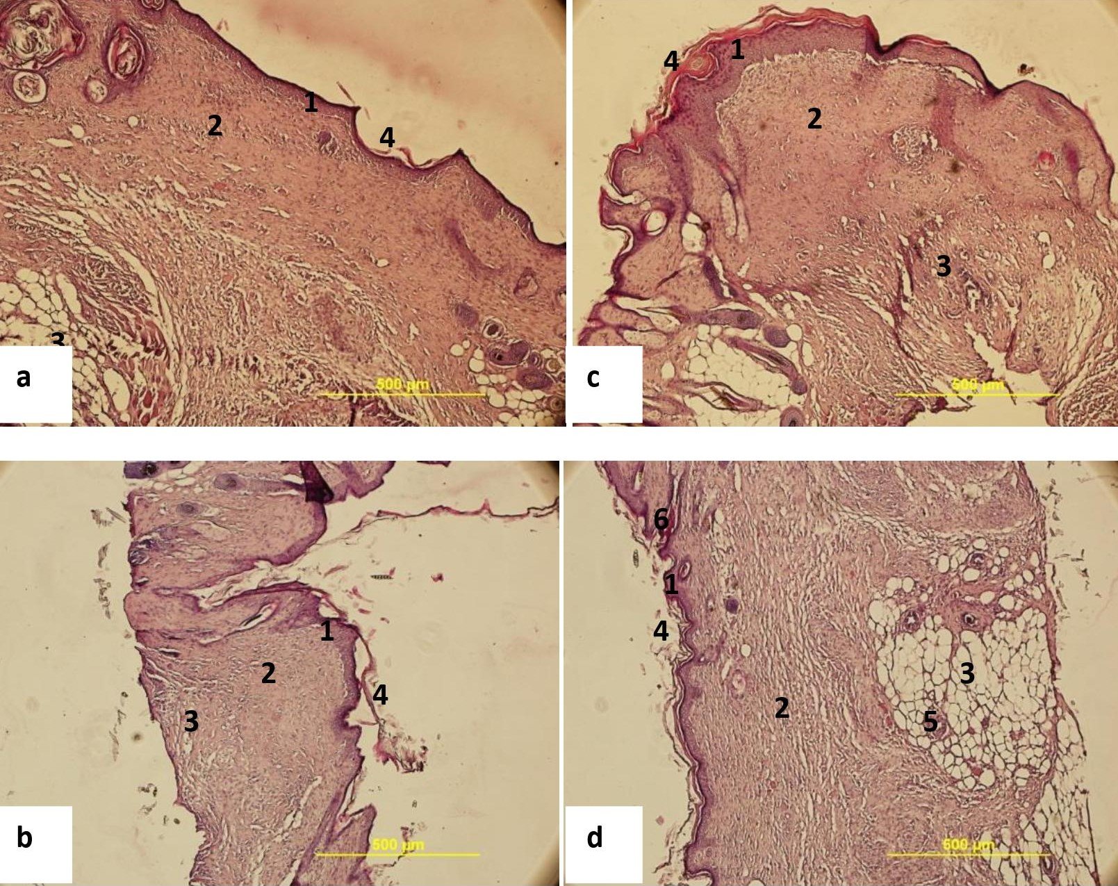
Fig. 3. Healing of skin wound on day 13. Hematoxylin and eosin staining. 1)epithelium, 2) derma, 3) subcutaneous adipose tissue, 4) crust, 5) gland ducts; 6) hair follicles.
Day 13
On day 13 after wound infliction and infection with pus, the appearance a focus of chronic inflammation in its central zone and the formation of the connective tissue scar in the derma were observed; no complete re-epithelization was seen (Fig.3, a). In the control group (wounds without pus contamination), practically complete re- epithelization and only minor inflammation were noted. The derma was enriched with small capillaries, collagen fibers were arranged in dense cords parallel to the epidermis, i.e. the formation of fibrous scar was observed. Scar formation was also noted in the subcutaneous tissue, collagen fibers were somewhat looser than in the derma, but adipose cells were almost absent (Fig.3, b). The wounds treated with levomecol were almost completely re-epithelized, but the epithelium detached from the derma. Inflammatory foci were seen in the derma. Subcutaneous adipose tissue was poorly developed, adipose cells were absent. Fibrous scar formed (Fig.3, c). Application of chitosan gel containing S.M.E.M.®, pronounced retraction of the wound edges, and its complete re-epitelization were noted (more pronounced that in groups 1 and 2); no epithelium detachment was observed. The structure of the derma was almost restored, no inflammatory foci were seen. Restoration of numerous gland ducts in the subcutaneous tissue and hair follicles in the derma was observed. The adipose tissue was well- developed. Partial restoration of muscle elements was noted somewhere under the wound. These findings attest to extremely high efficiency of the test composition in skin wound healing in mammals in vivo. The preparation not only stimulated regeneration i n the skin wound, but also promoted the formation of morphologically restored tissue in the injured area (Fig.3,d)
Thus, the test chitosan-based gel containing S.M.E.M.® (Signalling Molecules of the Extracellular Matrix) not only promotes tissue recovery in the injured area, but also arrests inflammation and limits the development of the infection process caused by pathogenic microflora of the pus. It was found that chitosan-based composition containing signaling molecule actively participates in the regeneration of skin wounds in mammals in vivo. It ensures practically complete recovery of the morphological structure of injured skin. This composition can be used for prevention of scar formation and healing of purulent skin wounds.